For half a century, biochemists have grappled with a stubborn enigma: why do certain proteins behave unpredictably when subjected to acidic environments? Recent findings now suggest that the answer lies not in the proteins themselves, but in the water molecules surrounding them. As acidity rises, water appears to migrate away from protein surfaces in ways scientists hadn't fully appreciated—a discovery that could reshape our understanding of cellular stress, drug design, and industrial bioprocessing.
The puzzle dates back to early observations in the 1970s, when researchers noticed that lowering pH altered protein structures in ways traditional models couldn't explain. Textbooks taught that acids donate protons, changing the charge on amino acid residues and destabilizing the folded shapes proteins depend on to function. Yet experimental data repeatedly showed deviations from these predictions, particularly in the intermediate pH range between 4.0 and 6.0. Some proteins collapsed faster than expected; others stubbornly retained their native folds. The inconsistency suggested a missing variable.
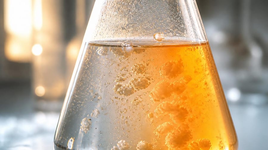
Water's Silent Role in Protein Stability
Proteins don't exist in isolation. They're bathed in water, and every fold, twist, and functional site depends on an intricate dance with H₂O. Hydration shells—layers of water tightly bound to protein surfaces—help maintain structural integrity. When pH drops, protons flood the environment. Until now, scientists focused on how those protons interact with the protein's charged groups. What they underestimated was the simultaneous disruption of hydration networks.
New biophysical studies using advanced spectroscopy and molecular dynamics simulations reveal that acidic conditions don't just protonate amino acids—they also strip away water molecules from specific protein regions. Hydrogen bonding patterns shift; water clusters that once stabilized loops and grooves dissipate. The protein, suddenly less cushioned, responds by collapsing inward or exposing hydrophobic patches that normally stay buried. This phenomenon appears most pronounced in proteins with large surface areas and shallow binding pockets.
Understanding how water exits the protein microenvironment under acidic stress opens new pathways for designing therapeutics that remain stable across pH gradients.
Implications for Drug Development and Biotechnology
The pharmaceutical industry has long wrestled with formulation challenges. Many injectable drugs—antibodies, insulin analogs, enzyme replacements—must remain stable in solutions that drift toward acidity during storage or in the body's acidic compartments, such as the stomach or endosomes. A protein therapeutic that unfolds prematurely loses its efficacy and may trigger immune reactions. By recognizing that water loss rather than simple protonation drives much of this instability, formulators can now design excipients—stabilizing agents—that preserve hydration shells even at low pH.
Industrial bioprocessing also stands to benefit. Fermentation tanks, protein purification columns, and enzyme reactors often operate under conditions where pH fluctuates. Engineers have relied on trial-and-error buffering strategies. With clearer insight into acid-driven dehydration, process designers can optimize buffer compositions and ionic strength to maintain the critical water layer around catalytic sites, improving yield and reducing costly batch failures.
How Scientists Uncovered the Mechanism
Breakthroughs in experimental technique made the discovery possible. Terahertz spectroscopy, which probes water's vibrational modes on picosecond timescales, allowed researchers to watch hydration shells disintegrate as pH dropped. Neutron scattering studies, sensitive to hydrogen atoms, mapped water density around protein surfaces at atomic resolution. Computational chemists then ran million-timestep simulations, tracking individual water molecules as protons entered the system.
Key observations included:
- Water molecules nearest acidic residues (aspartate, glutamate) detached first, within nanoseconds of protonation.
- Hydrophobic regions that normally repel water suddenly became dehydrated hotspots, destabilizing adjacent secondary structures.
- Proteins with intrinsically disordered regions showed heightened sensitivity, collapsing into compact, aggregation-prone forms.
- Buffers containing polyols (such as glycerol) or certain salts mitigated water loss by forming alternative hydrogen-bonding networks.
These findings converge on a unifying principle: acidity perturbs not just charge but the entire hydration landscape that proteins depend on.
Broader Impact on Cellular Biology
Inside living cells, pH varies by compartment. Lysosomes maintain a pH around 4.5 to activate digestive enzymes; the cytoplasm hovers near 7.2. Proteins trafficking between these zones must tolerate dramatic pH swings without misfolding. The new understanding suggests that cellular chaperones—proteins that help others fold—may also act as hydration managers, recruiting or releasing water to buffer pH-induced stress.
Cancer cells often acidify their surroundings to promote invasion and evade immune attack. Tumor-associated proteins exposed to this low-pH microenvironment may lose water and adopt aberrant conformations, affecting signaling pathways and drug responsiveness. Oncologists and biochemists are beginning to explore whether targeting hydration dynamics could offer a novel therapeutic angle, complementing traditional approaches that focus on inhibiting mutant enzymes or blocking receptors.
Comparing Old and New Models
| Aspect | Traditional View | Revised Understanding |
|---|---|---|
| Primary driver | Protonation of charged residues | Coupled protonation and water loss |
| Focus | Electrostatic repulsion, charge shielding | Hydration shell integrity, hydrogen-bond networks |
| Prediction accuracy | Moderate, especially at intermediate pH | Improved across pH 4.0–6.0 range |
| Intervention strategy | Buffering, ionic strength adjustment | Hydration stabilizers, polyol excipients |
Future Directions and Open Questions
While the core mechanism is now clearer, many questions remain. How do different amino acid sequences modulate water retention under acidic stress? Can synthetic polymers mimic natural chaperones to stabilize therapeutic proteins in harsh environments? What role does temperature play—does heat accelerate acid-driven dehydration, or do cold conditions trap water in place?
Researchers are also investigating whether similar dynamics govern protein behavior under alkaline conditions, where hydroxide ions might induce distinct hydration changes. Early data hint at asymmetries: bases may recruit water rather than expel it, leading to swelling and aggregation through a different pathway.
For bioengineers, the practical payoff is immediate. Next-generation protein designs may incorporate hydrophilic tags or surface mutations that anchor water molecules even at low pH. Formulation scientists are already testing novel excipient cocktails inspired by the hydration-preservation principle. And structural biologists are revisiting decades-old crystal structures, reinterpreting anomalous electron density as evidence of disrupted water networks rather than disordered loops.
This information does not replace advice from a qualified professional. Consult with biochemists, pharmaceutical scientists, or regulatory experts before applying these concepts in drug development or clinical settings.